What is Echo test and what are the techniques used for the echo test?
An echocardiogram uses ultrasound technology to determine the health of your heart’s structure and function. Cardiomyopathy and valve disease, for example, may be accurately diagnosed using an echo. Transthoracic and transesophageal echo exams are two examples of the many kinds of echo examinations available.
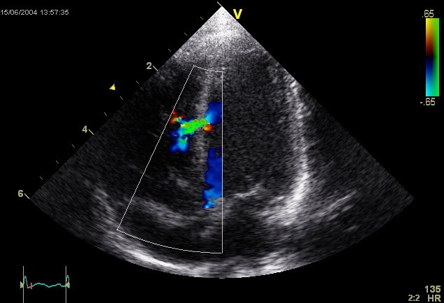
Graphic representation of your heart’s action, in the form of an echocardiogram (echo). The heart’s valves and chambers are photographed using ultrasound high-frequency sound waves delivered by a hand-held probe pressed against your chest. This aids in the evaluation of your heart’s pumping motion. Echocardiography, Doppler ultrasonography, and color Doppler methods are routinely used to examine blood flow through your heart’s valves. Get echo test in bangalore quickly at every hospital.
No radiation is used in the process of doing an echocardiogram. This distinguishes an echo from more minor radiation-intensive procedures like X-rays and CT scans.
A person carries out an echo test, but who is that person?
Your echo is performed by a specialist known as a cardiac sonographer. They know how to do echo testing and use the most up-to-date equipment. For example, they are trained to work in hospital rooms and catheterization laboratories. It is well known that Dr Pavan Rasalkar is an expert in this field. Get a famous cardiologist in banashankari easily at genuine fees.
What echocardiography methods are employed?
Echocardiography might take anywhere from 40 to 60 minutes. A TE might take up to 90 minutes to perform. There are a variety of methods for creating heart-shaped images. The optimal strategy for you relies on your medical condition and the demands of your doctor. The following techniques will be taught to you at the hospital:
- Ultrasound imaging in two dimensions: This is the most common method. The resulting 2D pictures are shown as “slices” when viewed on a computer screen. These slices may have been “stacked” to create a three-dimensional object in the past. Get heart care clinic banashankari for those who are suffering from heart issues.
- Ultrasound in three dimensions (3D): 3D imaging has become more efficient and helpful due to technological advances. New 3D methods allow you to see your heart in more detail, including how effectively it pumps blood. Your sonographer will be able to get a better view of your heart because of the 3D technology.
- Doppler echocardiography: This method may tell you how quickly and in what direction your blood is moving. Search for cardiologists dortor near me and get the results at your fingertips.
- Ultrasound using colour Doppler: Using a distinct hue for each flow direction, this approach illustrates your blood flow.
- The imagery of the stress response: This method demonstrates alterations in the way your heart muscle contracts. Heart disease may be detected in its earliest stages. Ablood pressure specialist doctor will help to know the blood pressure quickly within minutes.
- Contrast MRI/CT: Your healthcare practitioner injects a contrast agent into one of your veins. In the photographs, you can see the material, which may enable you to see the intricate features of your heart. The contrast agent may cause moderate allergic responses in certain patients, although this is rare.


